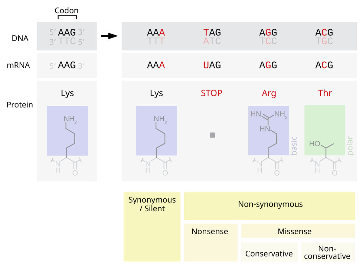
Which Protein Subunits Are Depicted In The Diagram
Then elf5b gdp and elf1a are released. A ribosome has two subunits.
 Dynamics Of Ribosomes And Release Factors During Translation
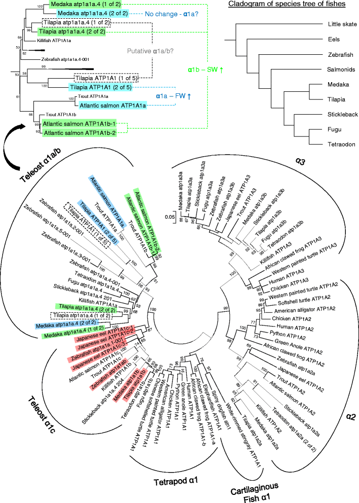
Three each of alpha and beta and 8 of little c again in vertebrates.

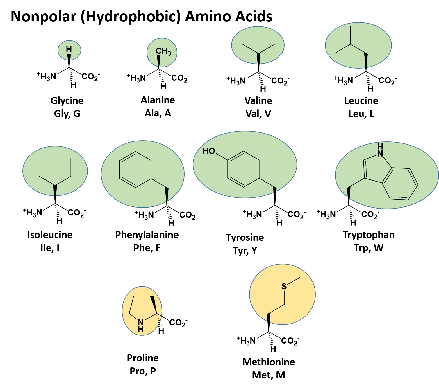
Which protein subunits are depicted in the diagram. Since this cap is present in the diagram the. Polypeptide chains generally contain both hydrophobic and hydrophilic residues. Much like detergent micelles proteins are most stable when their hydrophobic parts are buried while hydrophilic parts are on the surface exposed to water.
However the 40s subunit is much larger than the prokaryotic 30s subunit and contains ma. Complex i the most complex has 49 or 50. Each ribosome has two unequal subunits a large and a small subunit.
Correct the subunits present depend on whether the ribosome is eukaryotic 40s and 60s or prokaryotic 30s and 50s. The eukaryotic small ribosomal subunit is the smaller subunit of the eukaryotic 80s ribosomes with the other major component being the large ribosomal subunit. This accurately describes the polarity with which the message is read as well as the direction of protein synthesis.
They have combined sedimentation coefficient of 70s. 7 the 60s subunit will now combine with the 40s subunit resulting in release of most of the initiation factors. Primary secondary tertiary quaternary.
Both subunits contain many proteins and at least one large rrna. Select all that apply. The 40s and 60s names originate from the convention that ribosomal particles are denoted according to their sedimentation coefficients in svedberg units.
Elf5 gtp is now bound to elf1a in the a site. Proteins form by amino acids undergoing condensation reactions in which the amino acids lose one water molecule per reaction in order to attach to one another with a peptide. Protein structure is the three dimensional arrangement of atoms in an amino acid chain molecule.
The 7methylguanosine m7g cap is added to the 5 end of the premrnas in eukaryotes. Complex iv the oxidase has 13 in vertebrates. A single amino acid monomer may also be called a residue indicating a repeating unit of a polymer.
Proteins are polymers specifically polypeptides formed from sequences of amino acids the monomers of the polymer. Vertebrate complex iii has 11 subunits all different. 8 correct association of the 40s and 60s subunits induces hydrolysis of the gtp bound to elf5.
Many cellular enzymes are composed of subunits. The atp synthase has multiple subunits some of which are present in multiple copies within the same molecule. Different subunits belonging to the same protein plex different subunits belonging to the same protein plex often exhibit discordant expression levels and evolutionary properties figure s1 a mechanism of covalent substrate binding in the x ray fig 1 structure of the dhakdha plex a ribbon diagram of the dhak dimer the subunits are gray the n terminal.
It is structurally and functionally related to the 30s subunit of 70s prokaryotic ribosomes. Bacterial ribosomes are composed of two subunits of 30s and 50s sedimentation coefficient in sucrose. Part c which protein subunits are depicted in the diagram.
The same classes of interactions that contribute to the stability of tertiary protein structure also serve to stabilize the quaternary association of subunits namely ionic bonds hydrogen bonds hydrophobic bonds and disulfide bridges. The ribosome moves down the mrna in the 5 to 3 direction and synthesizes protein in the direction of amino terminus to carboxyl terminus. Ribosomes and protein synthesis.


Solved 7 Hemoglobin Pictured Below And To The Left Con
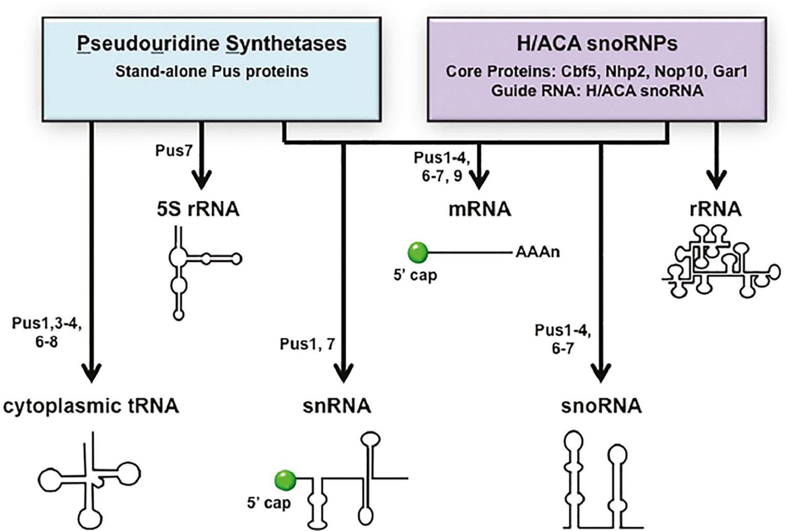 Frontiers Trna Processing And Subcellular Trafficking
 Proteinsplus A Web Portal For Structure Analysis Of
 Insights Into The Base Pairing Preferences Of 8 Oxoguanosine
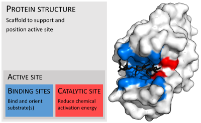 Active Site Wikipedia
 Elongation Of Polypeptide Chain Expii
 Figure 7 From The E Coli Btucd Structure A Framework For
 A Potential Architecture Of The Ino80 Complex The 15

 Architecture Of The Heteromeric Glua1 2 Ampa Receptor In
 Binding Assays Of Variant Coat Proteins And Dec To Probe The
 Flexible Selection Of Diversified Na K Atpase A
 N Degron And C Degron Pathways Of Protein Degradation Pnas
 Semaphorin3a Signaling Is Dispensable For Motor Axon
 The Biological Building Blocks Cancerquest
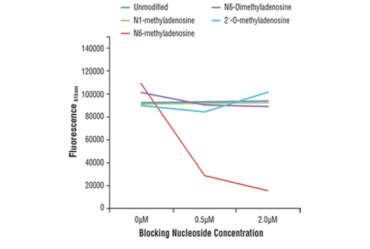 Cst N6 Methyladenosine M6a D9d9w Rabbit Mab
 Vibratory Urticaria Associated With A Missense Variant In
 Point Mutation Wikipedia
 Vibratory Urticaria Associated With A Missense Variant In
 Mthsp70 Antibody Ma3 028
 Schematic Diagram Depicting The Nox2 Containing Nadph
 Solved 7 Hemoglobin Pictured Below And To The Left Con

Translation Study Guide
0 Response to "Which Protein Subunits Are Depicted In The Diagram"
Post a Comment