Diagram Of Back Muscles
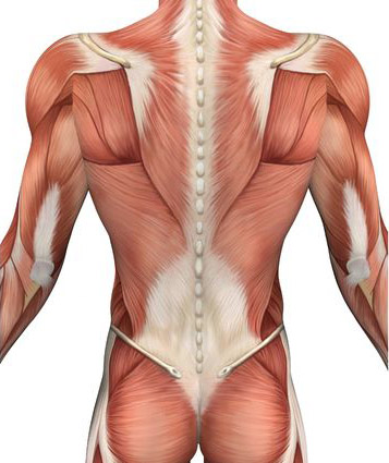
Three types of back muscles that help the spine function are extensors flexors and obliques. 7 muscle diagram lower back and moreanatomy muscles lower back hip muscle anatomy of lower back and buttocks muscle chart lower back muscle diagram lower back muscle human diagram chart.
 Superficial And Deep Back Muscles Examination Of The
The extensor muscles are attached to the back of the spine and include the large muscles in the lower back which help hold up the spine and gluteal muscles.
Diagram of back muscles. All these muscles are therefore associated with movements of the upper limb. The intrinsic back muscles. The clavicle scapula and humerus.
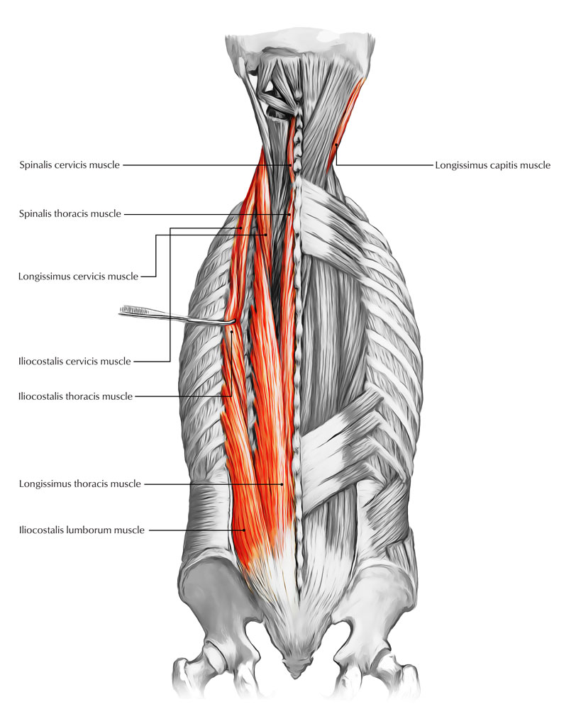
Names and diagram superficial muscle group. The spinal cord and its nerves are the means by which the body and brain communicate with one another. The deep muscle group can be subdivided into four smaller groups muscles.
Organs found in the back. They originate from the vertebral column and attach to the bones of the shoulder. Start now for free.
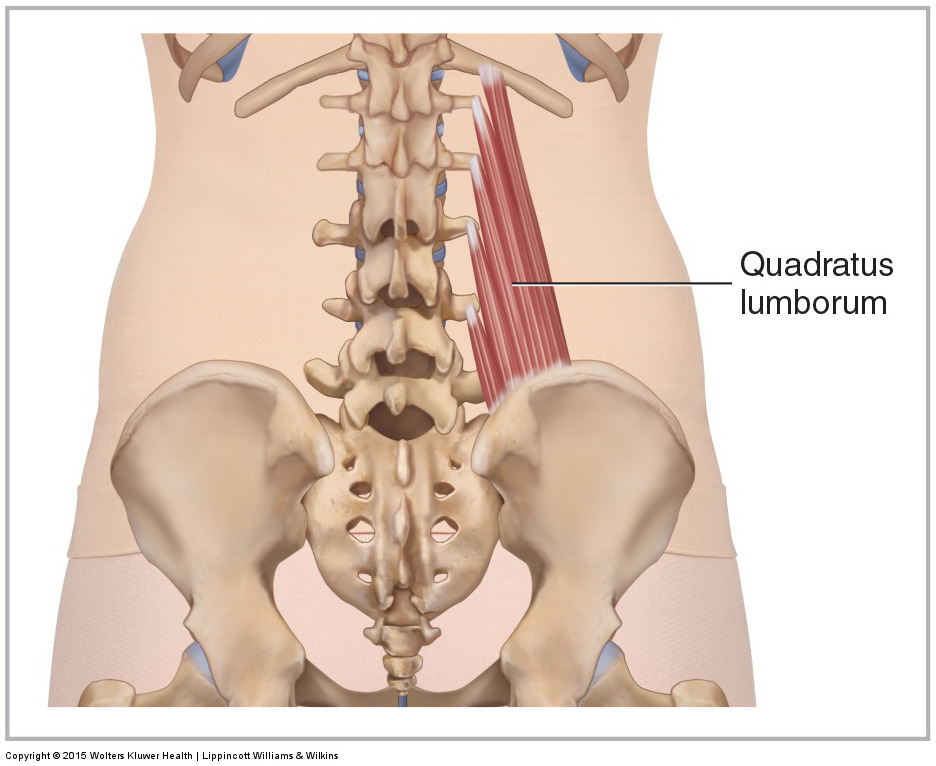
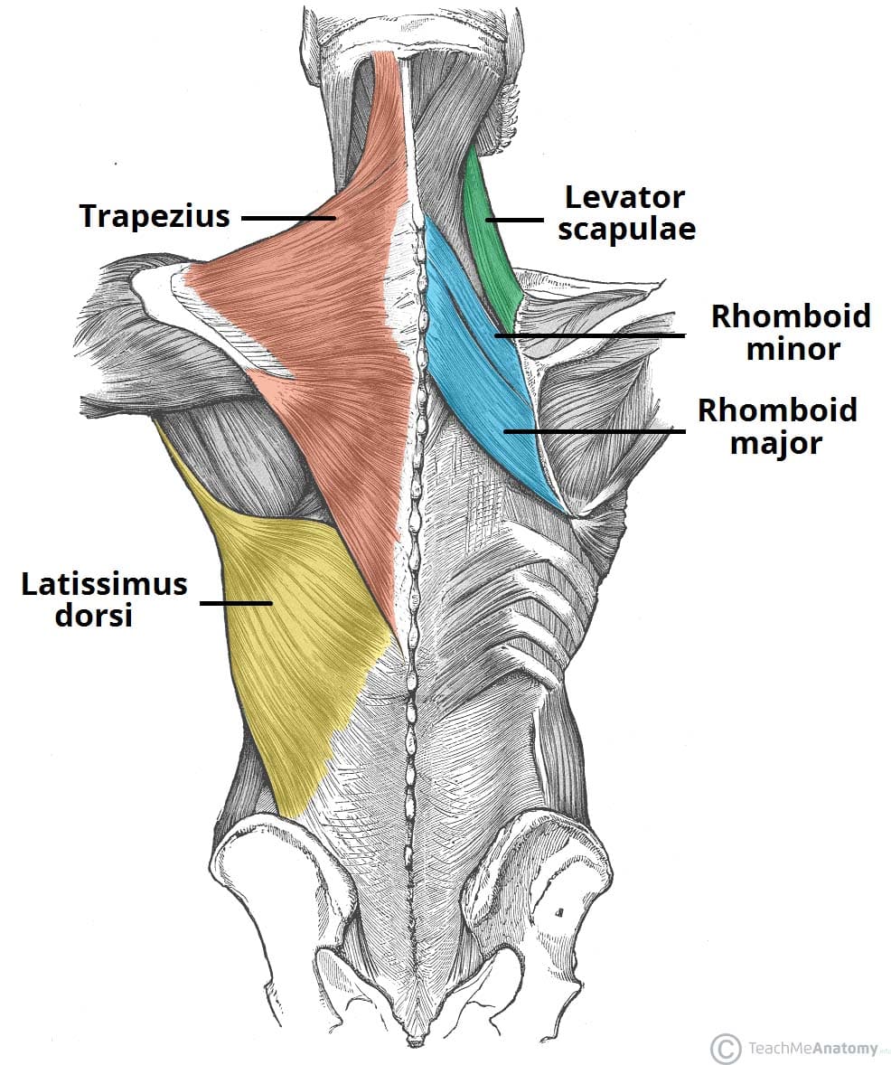
Lower back and superficial muscles. Meanwhile the flexor muscles are attached to the front of the spine and include the abdominal muscles. Both muscles are innervated by the dorsal scapular nerve c4 to c5 which is a branch of the brachial plexus.
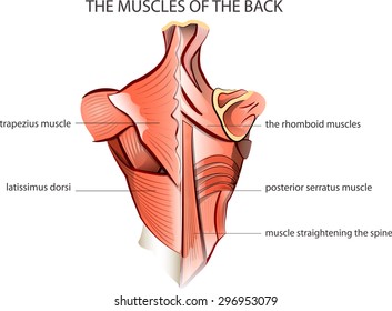
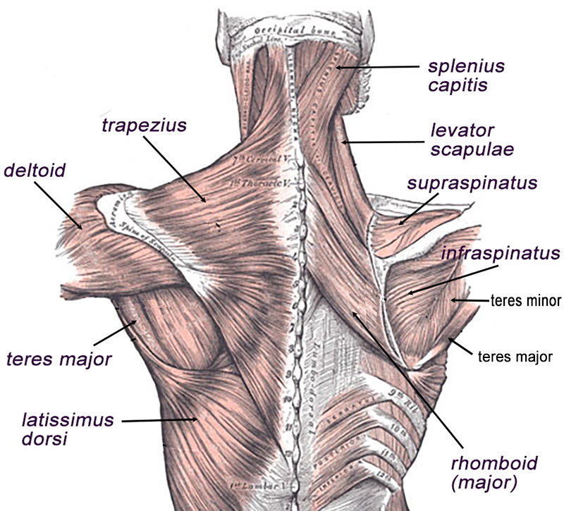
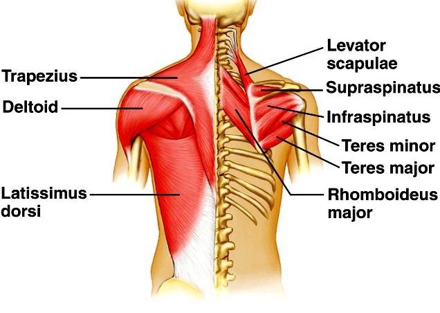
Part of the teachme series. The extensor muscles are attached to back of the spine and enable standing and lifting objects. The muscles in this group are the trapezius latissimus dorsi levator scapulae and the rhomboids.
This information is intended for medical education and. The trapezius or trapezoid muscles are two paired muscles. Oblique muscles are attached to the sides of the spine.
The muscles of the lower back help stabilize rotate flex and extend the spinal column which is a bony tower of 24 vertebrae that gives the body structure and houses the spinal cord. Diagram of back muscles labeled image via humanbodyanatomyco diagram of back muscles blank image via humanbodyanatomyco. These muscles include the large paired muscles in the lower back called erector spinae which help hold up the spine and gluteal muscles.
The medical information on this site is provided as an information resource only and is not to be used or relied on for any diagnostic or treatment purposes. Rhomboid minor has its origin on the spinous process of the c6 to t7 vertebrae and also inserts on the medial border of the scapula above the spine of the scapula.
 Back Muscles Overview Stretching Strengthening Ex How
 Back Muscles 28 Major Muscles Of The Back Earth S Lab
![]() Back Muscles 28 Major Muscles Of The Back Earth S Lab
Back Muscles 28 Major Muscles Of The Back Earth S Lab
 Back Muscles Diagram Quizlet
 Diagram Back Muscles And Lower Back Muscles Diagram Lower
 Mini Poster Shoulder And Back Muscles Anatomy 34x24 Cm Med
 Muscles Of The Lumbar Spine Of The Trunk

 Muscles Of The Human Back Science Of Anatomy
 Muscles Of The Back Teachmeanatomy
 Back Of Neck Muscles Diagram Wiring Diagrams
 Back Muscle Anatomy Images Stock Photos Vectors
 Muscles Of The Back
![]() Anatomy Of The Back Spine And Back Muscles Kenhub
Anatomy Of The Back Spine And Back Muscles Kenhub
![]() Back Muscles Anatomy And Functions Kenhub
Back Muscles Anatomy And Functions Kenhub

 Back Muscles Quiz By Jeewonprk
 34 Always Up To Date Human Anatomy Back
 Pin By Reyman Panganiban On Anatomy In 2019 Shoulder


 A P I Quiz Back Muscles Diagram Quizlet
 Major Posterior Muscles Anatomy Move Your Body Muscle
 What Are The Causes Of Low Back Muscle Spasming
 Upper Back Muscles Diagram Quizlet
 Back Muscles Attachments Nerve Supply Action Anatomy Info
 Anatomy Of The Spine And Back
0 Response to "Diagram Of Back Muscles"
Post a Comment