Mouse Heart Diagram
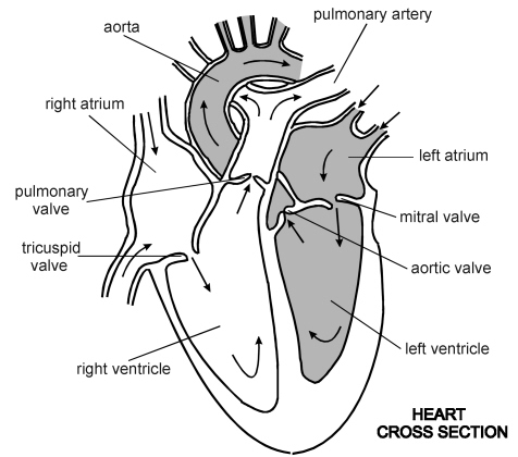
Blood vessels of the neck. The heart has four chambers.
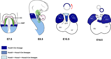
Patterning Of Mouse Heart Development A Hierarchical
Blood vessels of axilla.

Mouse heart diagram. Mouse anatomy rat and mouse anatomy comparative anatomy of the mouse and rat. Under surface or right hind foot. Upper surface of right hind foot.
Selecting or hovering over a box will highlight each area in the diagram. In this interactive you can label parts of the human heart. A rabbit has 205bpm.
Label these on the diagram. The right ventricle receives blood from the right atrium and pumps it to the lungs where it is loaded with oxygen. Blood vessels of right hind limb.
Superficial dissection of the neck and thorax. A blue whales heart beats six times a minute. Blood flows from the right atrium to the right ventricle via the tricuspid valve.
Insert the 30 gauge needle from the tubing with saline10 formalin solution into the apex of the left ventricle. Blood from the posterior portion of the body enters the right atrium of the heart through the inferior vena cava and the superior vena cava. A horse has 38bpm.
Drag and drop the text labels onto the boxes next to the diagram. Label each on the diagram. Be careful to not pierce the heart.
Using clamp scissors grab at the base of the sternum cut through the ribcage and lift to expose the heart. A diagram displaying the blood flow in the heart of a late prenatal mouse. The heartbeat of a canary occurs 17 times in a second.
Trace the flow of blood inside the heart. The right atrium receives blood from the veins and pumps it to the right ventricle. In mice the cardiac veins run on the surface of the heart within the subepicardium draining the myocardium of the left and the right ventricles as well as the left atrium.
However some abnormal veins have been described in various malformations of the human heart uemura et al. The average heart rate for a man is 72bpm. Drag and drop the text labels onto the boxes next to the heart diagram.
In this interactive you can label parts of the human heart. The left atrium receives oxygenated blood from the lungs and pumps it to the left ventricle. A color atlas and text provides detailed comparative anatomical information for those who work with mice and rats in animal research.
Transfer the mouse to the procedural stageplatform. Oxygenated blood from the placenta that enters the right atrium is shunted through the inter atrial septa via the foramen ovale to the left atrium and through the left atrioventricular. Male organs deflected to left to show blood supply.
Blood supply of testis. Blood supply of left kidney and suprarenal gland. Blood vessels of lower right hind limb.
Skin removed from dorsal surface of right hind limb.
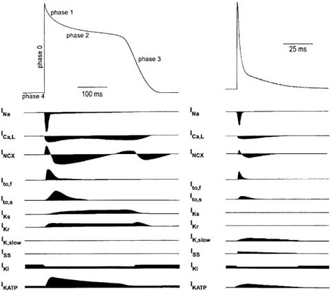 Molecular Diversity Of Ion Channels In The Mouse Heart

 Cardiac Lymphatic Drainage Of An Adult Mouse Heart
 Human Mouse Rat Fabp3 H Fabp Antibody Mab1678 R D Systems
 Locally Expressed Igf1 Propeptide Improves Mouse Heart
 Mouse Heart Shape Stock Photo 103647023 Alamy
![]() Red Computer Mouse With Wire In Diagram Shape On White
Red Computer Mouse With Wire In Diagram Shape On White

 Mouse Heart By Josh Gomez On Prezi
 Figure 2 From Cardiac And Coronary Function In The

 Amazon Com I Ll Love You Forever Heart Shaped Mouse Pad
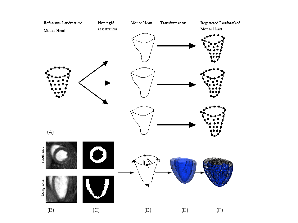 Study Of The Murine Cardiac Mechanical Function Using
 The Local Microenvironment Limits The Regenerative Potential
 Lowering Body Weight In Obese Mice With Diastolic Heart
 Depressed Heart Rate Variability And Arterial Baroreflex In
 Rat Heart Cartoon Vector Photo Free Trial Bigstock
 Building And Re Building The Heart By Cardiomyocyte
 Flow Diagram Of The Procedures For Mouse Dissection And
 Anatomy And Physiology Of Animals Cardiovascular System The
 A Diagram Displaying The Blood Flow In The Heart Of A Late
Plos One Pressure Overload Induced Angiotensin Mediated

0 Response to "Mouse Heart Diagram"
Post a Comment