The Diagram Below Shows A Bacterial Replication Fork And Its Principal Proteins
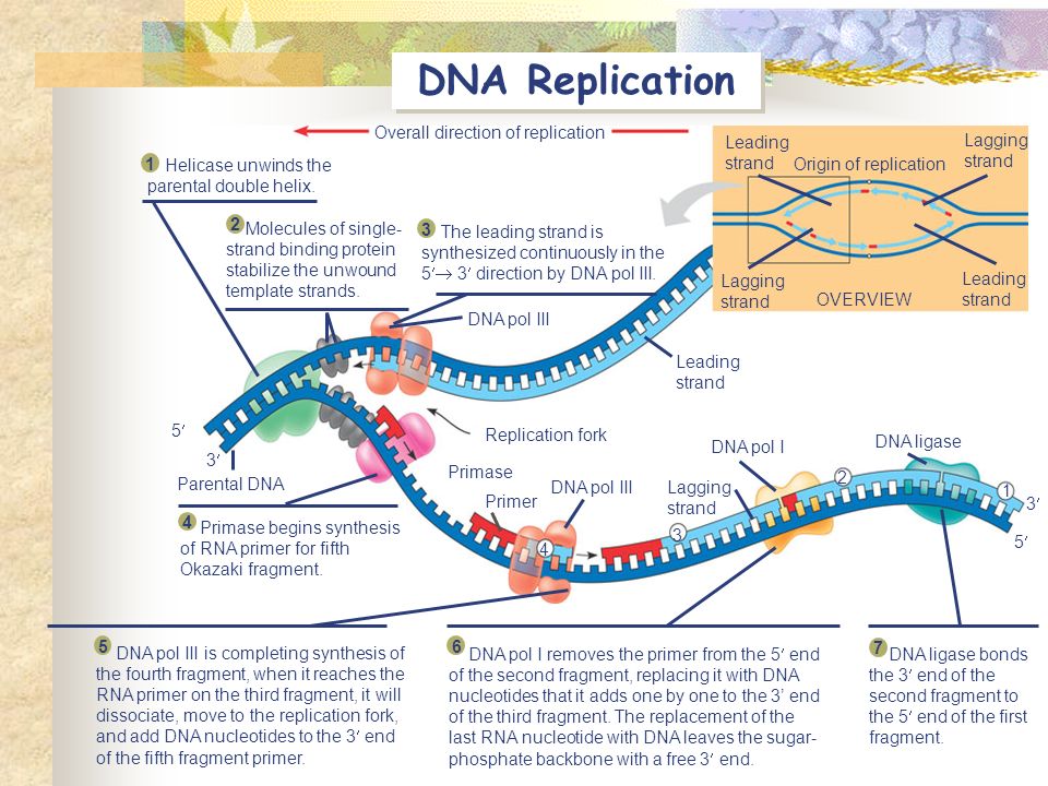
The diagram below shows a bacterial replication fork and its principal proteins. The diagram below shows a bacterial replication fork and its principal proteins.
The origin of replication is indicated by the black dots on the parental strands.

The diagram below shows a bacterial replication fork and its principal proteins. Answers the diagram below shows a bacterial replication fork and its principal proteins. The diagram below shows a replication bubble with synthesis of the leading and lagging strands on both sides of the bubble. Answers antibiotics free full text.
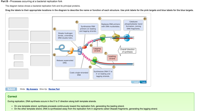
Use pink labels for the pink targets and blue labels for the blue targets. The diagram below shows a bacterial replication fork and its principal proteins. The parental dna is shown in dark blue the newly synthesized dna is light blue and the rna primers associated with each strand are red.
Use pink labels for the pink targets and blue labels for the blue targets. The diagram below shows a bacterial replication fork and its principal proteins. Use pink labels for the pink targets and blue labels for the blue targets.
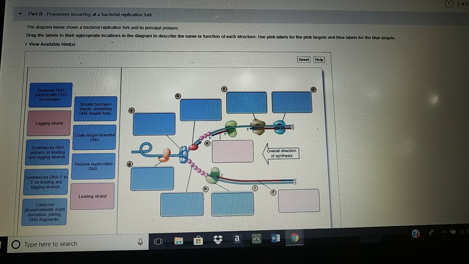
The diagram below shows a bacterial replication fork and its principal proteins. Part b processes occurring at a bacterial replication fork the diagram below from biol 4103 at university of houston. Drag the labels to their appropriate locations in the diagram to describe the name or function of each structure.
Use pink labels for the pink targets and blue labels for the blue targets. Drag the labels to their appropriate locations in the diagram to. The diagram below shows a bacterial replication fork and its principal proteins.
Shows a bacterial replication fork and its principal proteins. Drag the labels to their appropriate locations in the diagram to describe the name or function of each structure. Drag the labels to their appropriate locations in the diagram to describe the name or function of each structure.
Mastering biology exam 3. The diagram below shows a bacterial replication fork and its principal proteins. Use pink labels for the pink targets and blue labels for the blue targets.
Drag the labels to their appropriate locations in the diagram to describe the name. Drag the labels to their appropriate locations in the diagram to describe the name or function of each structure. Drag the labels to their appropriate locations in the diagram to describe the name or function of each structure.
 Mastering Biology Chapter 16 Rhs Homework
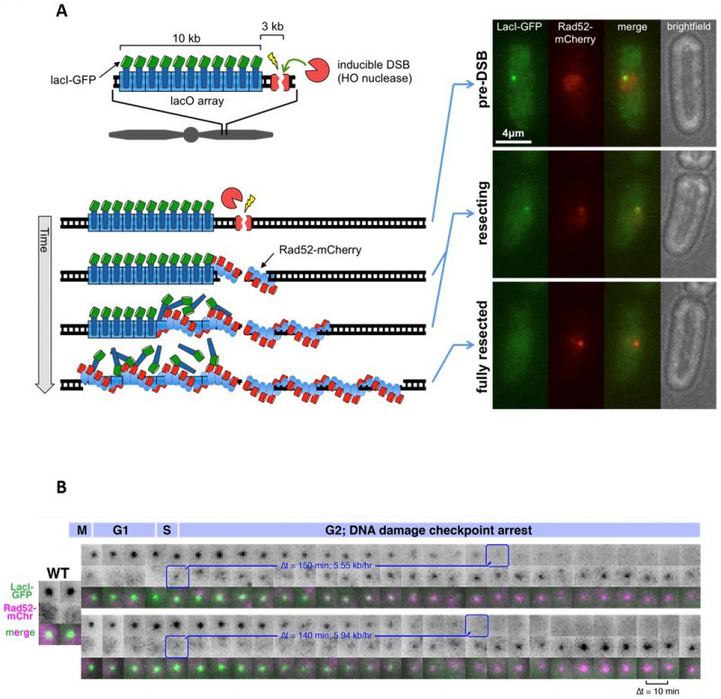
 Dynamics Of Dna Replication In A Eukaryotic Cell Pnas
 Mastering Biology Chapter 16 Rhs Homework
 Functions Of Multiple Clamp And Clamp Loader Complexes In

 The Diagram Below Shows A Bacterial Replic Clutch Prep
 The Diagram Below Shows A Bacterial Replic Clutch Prep
 Forging Ahead Through Darkness Pcna Still The Principal
 Mastering Biology Chapter 16 Rhs Homework
 Mastering Biology Chapter 16 Rhs Homework
 What Is Dna Replication Facts Yourgenome Org
 The Initial Response Of A Eukaryotic Replisome To Dna Damage
 Modern Dna Science And Its Applications Book Chapter
 Mastering Biology Chapter 16 Rhs Homework
 The Diagram Below Shows A Bacterial Replic Clutch Prep
 Stalled Replication Forks Generate A Distinct Mutational
 Cell Boundary Confinement Sets The Size And Position Of The
 Dna Replication Microbiology
 Mastering Biology Chapter 16 Rhs Homework
 Interactions Between Helicase And Primase Are Crucial For
Chapter 16 Homework In The Accompanying Image A Nucleotide
The Lactococcus Lactis Plasmidome Much Learnt Yet Still
 Honors Biology Ch 12 Molecular Genetics Ppt Video Online
 Primase Is Required For Helicase Activity And Helicase
 Multiscale Structuring Of The E Coli Chromosome By Nucleoid
 Dna Replication Microbiology
0 Response to "The Diagram Below Shows A Bacterial Replication Fork And Its Principal Proteins"
Post a Comment