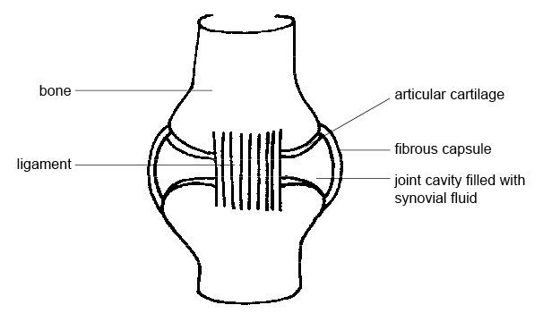
The Diagram Shows A Frontal Section Of The Hip Joint
A it is the fluid secreted by the knee joint that stabilize the joint bit is the partial fibrous capsule of the knee joint cit is the lateral ligaments of the knee that prevent hyper extension d it is articular cartilage that prevents the knee from rotating. Identity its major structural elements by using the letters key.
 Anatomy And Physiology Of Animals The Skeleton Test Yourself
Identify its major structural elements by using the key letters.

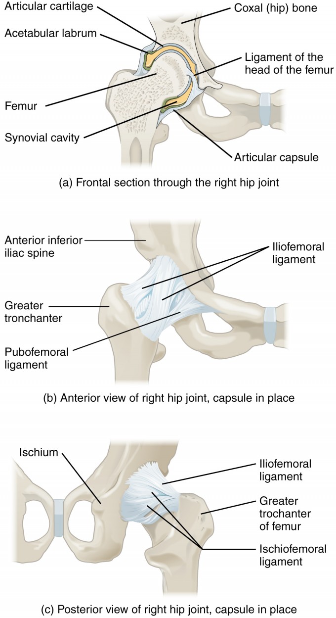
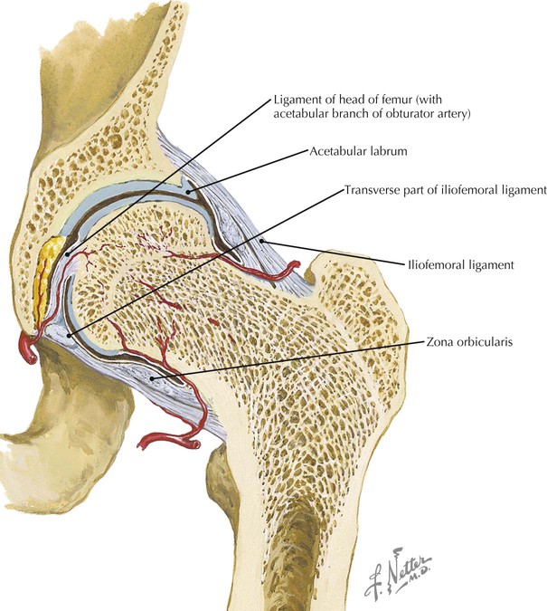
The diagram shows a frontal section of the hip joint. Posterior half viewed from in front. The diagram shows a frontal section of the hip joint. The menisci review sheet 13 and ligaments and tendons crossing joint 7.
The diagram on the right shows a cross section of the hip. The diagram shows a frontal section of the hip joint. 84 name two important factors that contribute to the stability of the hip joint.
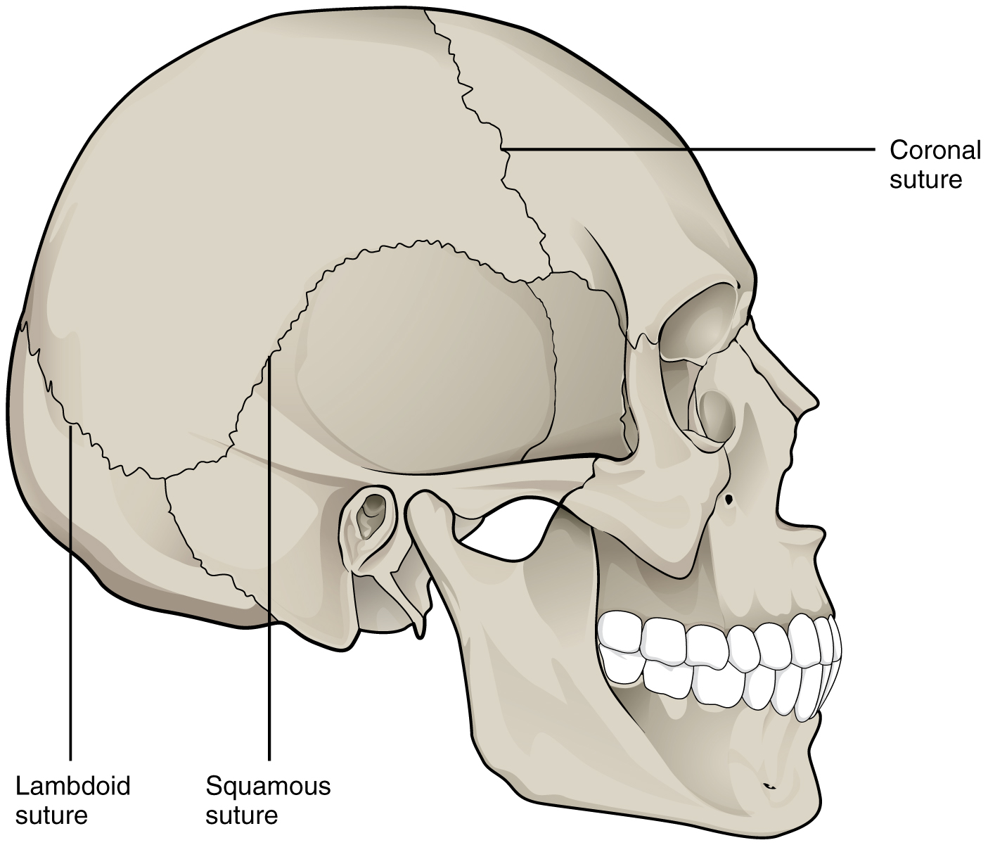
Head of femur f. The diagram on the left is a back view of the hip joint showing the thigh bone femur going into the pelvis bone held together by ligaments. Describe how the structure of the temporomandibular.
Deep socket for femur and strongly reinforced articular capsule name two important factors that contribute to the stability of the knee. Ligament of the bead of the femur the shoulder joint is built for mobility. Weight bearing stresses on the hip during walking can be 5 times a persons body weight.
The hip joint is one of the largest joints in the body and is a major weight bearing joint. Hip anatomy function and common problems. The diagram shows a frontal section of the hip joint.
The lacrimal bone the smallest and most fragile bone of the face is situated at the front part of the medial wall of the orbit. As you can see the top of the femur is shaped like a ball and the concave cavity of the pelvis is shaped like a socket. A thin scalelike bone roughly resembling a fingernail in size and shape at the anterior part of the medial wall of the orbit articulating with the frontal and ethmoidal bones and the maxilla and inferior nasal concha.
On the lateral aspect of the hip bone articulates with the head of the femur toarticulates with the head of the femur to form the hip joint th ili i hi d p bi j i t fthe ilium ishium and pubis join to form the acetabulum 33 8 from. Identify its major structural elements by using the key letters. The joint surfaces have been somewhat pulled apart.
Head of femur e. A healthy hip can support your weight and allow you to move without pain. The diagram shows a frontal section of the hip joint.
 Bones Of The Lower Limb Anatomy And Physiology I
 Anatomy Of Selected Synovial Joints Anatomy And Physiology I
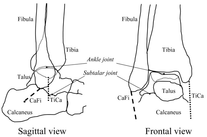
 Anatomy Of Lower Extremity
 Cross Sectional View Of The Normal Hip Joint Download
 Groin Injury Introduction And Frequency Functional Anatomy
 Pelvic Girdle Femur Sacroiliac Joint And Hip Joint
![]() Leg And Knee Anatomy Bones Muscles Soft Tissues Kenhub
Leg And Knee Anatomy Bones Muscles Soft Tissues Kenhub
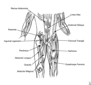
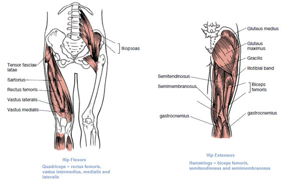 Muscles That Move The Leg

 Clinical Anatomy Of The Elbow And Shoulder Reumatologia
 Bones Of The Lower Limb Anatomy And Physiology I
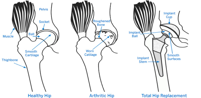
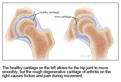 Treating Hip Arthritis Mu Health Care
 Frontal And Sagittal Views Of The Adult Spinal Column The

 Labeled Inner Structure Of A Hip Joint Exam 2 Joints Of The
 9 1 Classification Of Joints Anatomy And Physiology
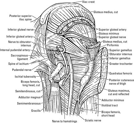 Nerves Of The Hip And Thigh Dummies
Snapping Hip Orthoinfo Aaos
 Lower Limb Radiology Key
Frontal Section Through Hip Joint Clipart Etc
 How Do I Know If I Need A Hip Replacement Hss Orthopedics
 Biomechanics Of Femoral Neck Fractures In Runners Lower


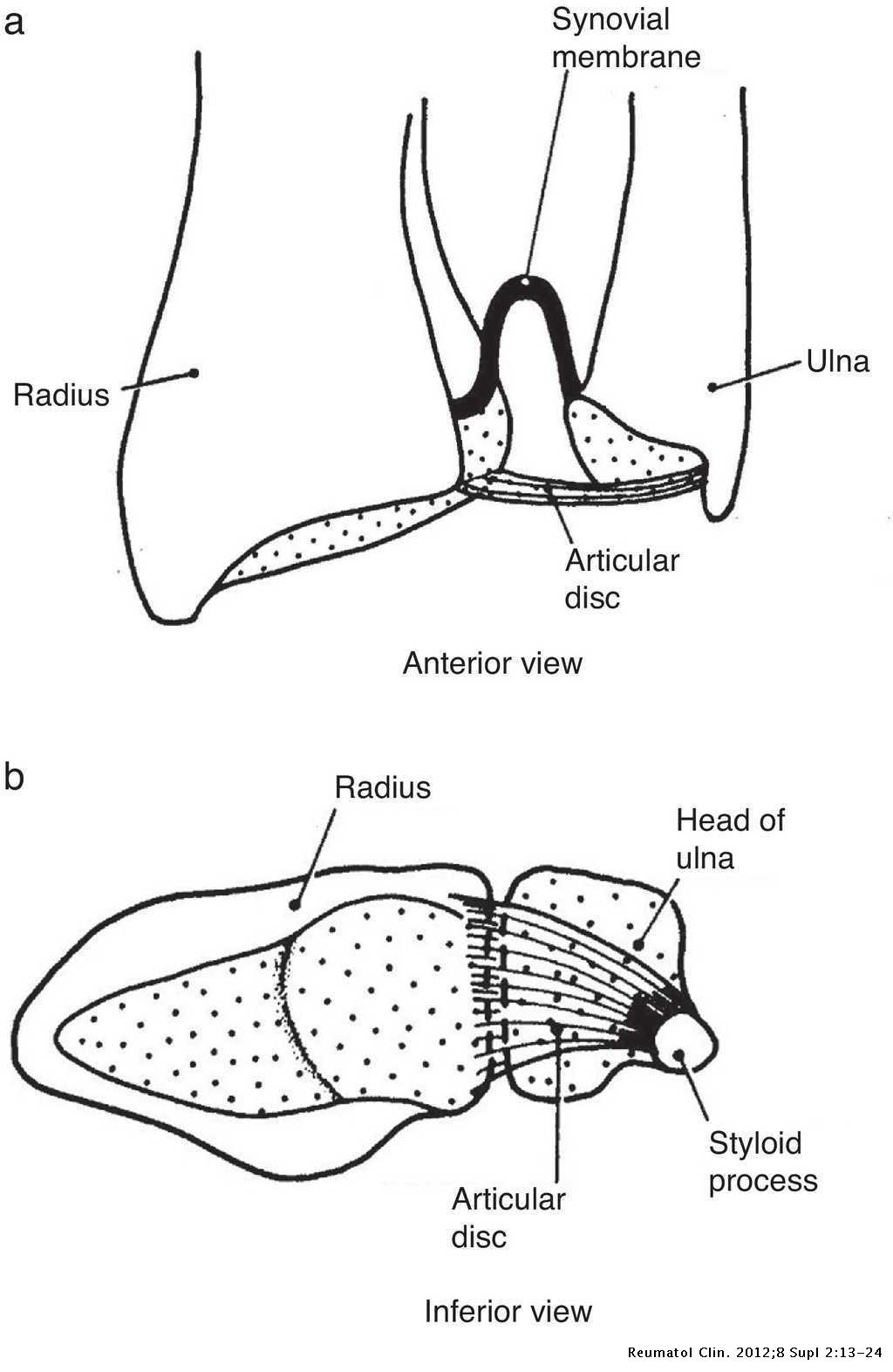 Clinical Anatomy Of The Elbow And Shoulder Reumatologia

 Frontal Section Through Hip Joint Clipart Etc
 Biomechanics Of The Natural Arthritic And Replaced Human
0 Response to "The Diagram Shows A Frontal Section Of The Hip Joint"
Post a Comment