Identify The Structures Labeled A B And C In The Diagram Of A Sarcomere Above
Identify the structures labeled a b and c in the diagram of a sarcomere above. Indicated by figure a in model 3.
 Skeletal Muscle Anatomy And Physiology I
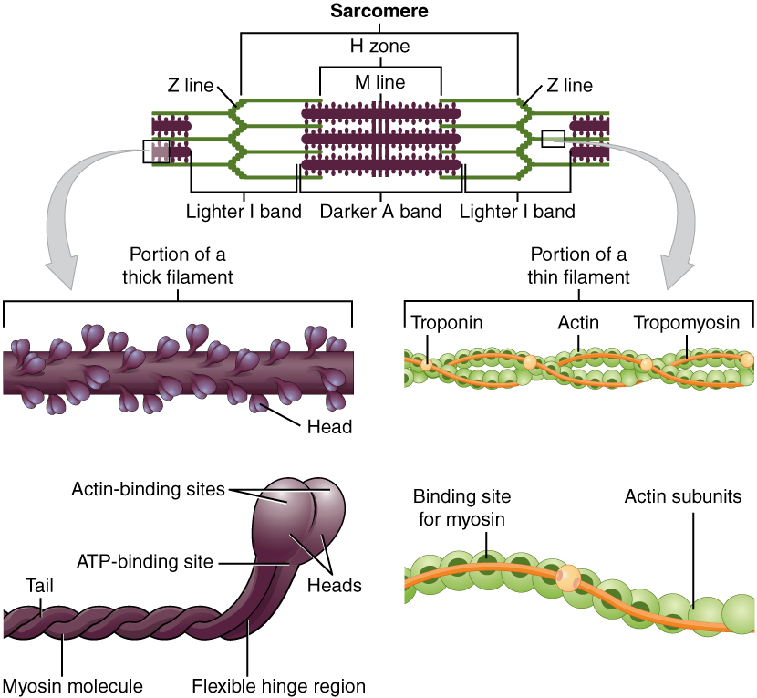
B myosin filament.

Identify the structures labeled a b and c in the diagram of a sarcomere above. Draw a vertical line and label it a. A actin filament. A at the beginning of a contraction.
In the diagram below draw three vertical lines showing the locations within a sarcomere of the cross sections indicated by figures a b and c. C in sarcomere 3 identify the location within the sarcomere of the cross section indicated by figure c in model 3. Calcium ions are released from the sarcoplasmic reticulum into the cytosol.
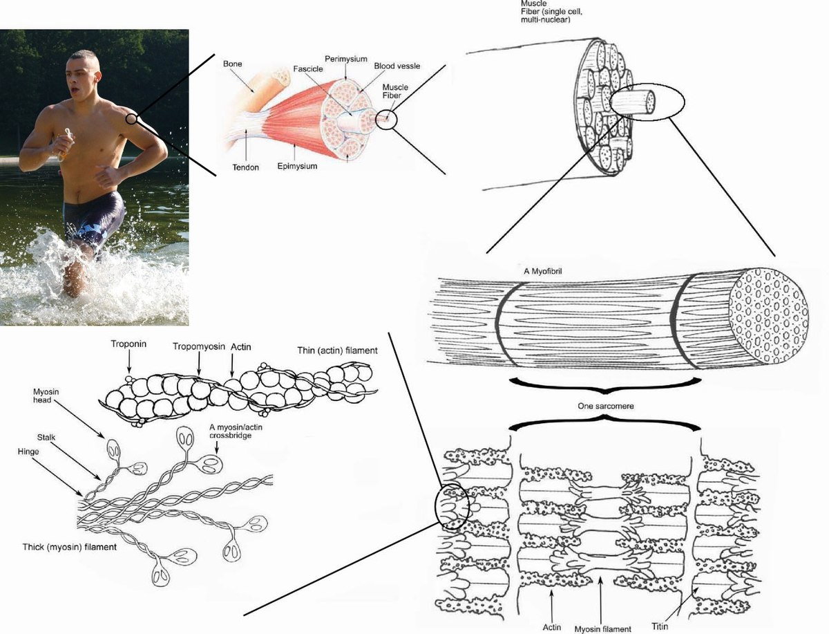
Section ii the organization structure and function of muscle the fundamental repeat unit within muscle that is responsible for contraction is the sarcomere. Locate the major hormone producing structures. Label the thick and thin filaments in figs.
Label each of the lines. C titin the sliding filament theory states that during muscle contraction. B in response to acetylcholine binding to ca2 release channels.
D after the contraction ends. This is a quiz called sarcomere labeling just point and click to play this. Students teachers and rockstars alike all come here to create and learn.
Label the thick and thin filaments in figs. A b and c above. Label the thick and thin filaments in figs.
The diagrams in model 3 are cross sections of a sarcomere that show the filaments at various locations within a sarcomere. Purposegames lets you create and play games. C by active transport using ca2 pumps in the sr membrane.
In the diagram below draw three vertical lines showing the locations within a sarcomere of the cross sections indicated by figures a b and c. A b and c above. The diagrams in model 3 are cross sections of a sarcomere that show the filaments at various locations within a sarcomere.
Play this quiz called label the sarcomere and show off your skills. B in sarcomere 2 identify the location within the sarcomere of the cross section indicated by figure b in model 3. Draw a vertical line and label it c.
Purposegames lets you create and play games. The sarcomere consists of a bundle of myosin containing thick filaments flanked and interdigitated with bundles of actin containing thin filaments fig. Various locations within a sarcomere.
Identify major abdominal arteries. A b and c above. In the diagram below draw three vertical lines showing the locations within a.
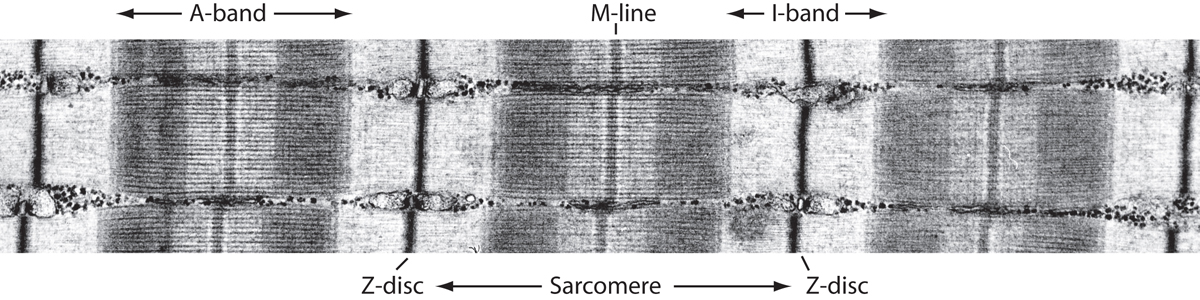
The light dependent reactions occur in the thylakoids. Which of the figures a b or c represents a cross section in the h zone. Students teachers and rockstars alike all come here.
E all of these answers are correct. The light independent reactions occur in the stroma. Draw a vertical line and label it b.

 Sarcomeric Organization And Mybp C A Cardiac Muscle
 Myosin Binding Protein C Corrects An Intrinsic Inhomogeneity
 Force Generation By Skeletal Muscle Is Controlled By
 Sliding Filament Model Of Contraction Biology For Majors Ii
 Muscles
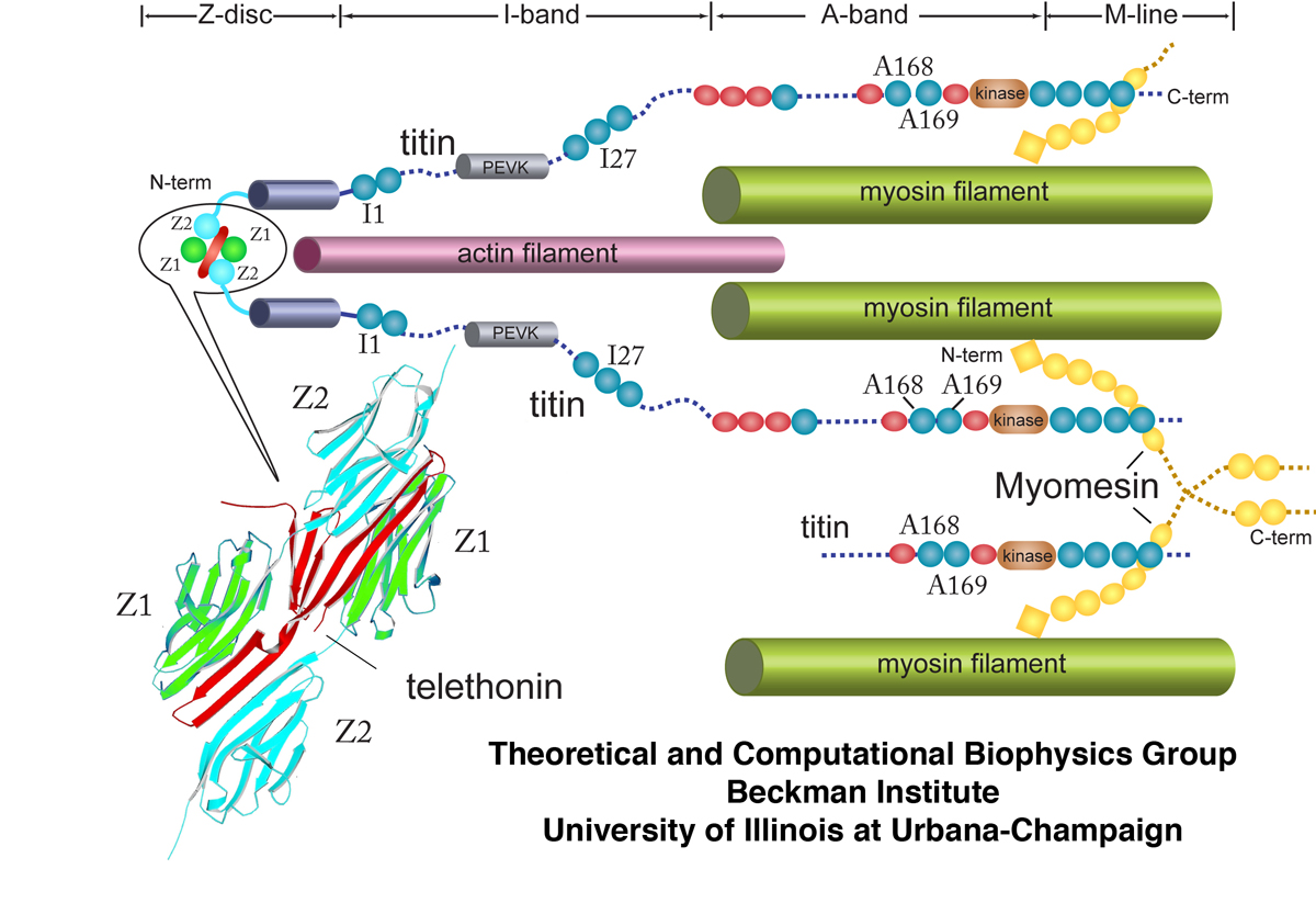
 Titin Wikipedia
 Sarcomere Length Tension Relationship Video Khan Academy
 10 2 Skeletal Muscle Anatomy And Physiology
 Sliding Filament Theory Wikipedia
 The Vertebrate Muscle Z Disc Sarcomere Anchor For Structure
 Types Of Muscle Tissue And Fibers Biology For Majors Ii
 The Sarcomeric Cytoskeleton From Molecules To Motion

 Skeletal Muscle Wikipedia
Schematics Of Sarcomere And M Band Structure Derived From
 Muscle Contraction And Locomotion Boundless Biology
 Muscle Tissue Junqueira S Basic Histology Text And Atlas
 Force Generation Via B Cardiac Myosin Titin And A Actinin
A Stochastic Simulation Of Skeletal Muscle Calcium

 The Sarcomere And Sliding Filaments In Muscular Contraction
 Myosin Binding Protein C Activates Thin Filaments And
0 Response to "Identify The Structures Labeled A B And C In The Diagram Of A Sarcomere Above"
Post a Comment