Integumentary System Diagram To Label
Epidermis the epidermis is the outer layer of the skin and is formed of five sub layers. Labeling of integumentary system.
 Integumentarysystemidentification Docx Integumentary
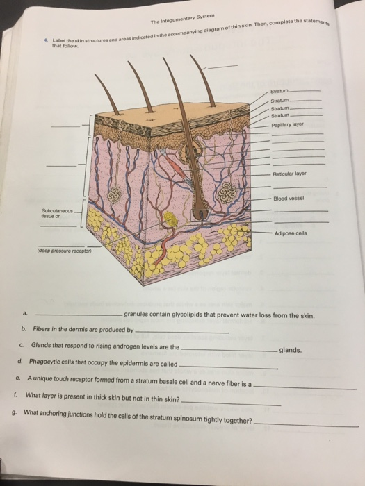
Glands that respond to rising androgen levels are the glands.

Integumentary system diagram to label. Well being the largest organ in the human body skin anatomy is certainly an important part of the integumentary system. Label the integumentary system. Melanocytes are the epidermal cells and are responsible for the synthesis of melanin pigment which in turn renders coloration to the skin.
Just pick an audience or yourself and itll end up in their incoming play queue. Arrector pili muscle identify the muscle. Skin and hair provide protection from harmful ultraviolet radiation and the skin guards against sunburn.
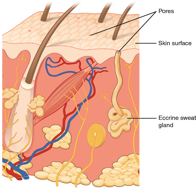
The layer under the stratum corneum in the epidermis in the skin of palms and soles. From outer to inner the layers are named the. But which of them are first to spring to your mind.
Students will add the provided labels to the diagram of the urinary system and then write a one sentence description of the function for each item. Granules extruded from the keratinocytes prevent water loss by diffusion through the epidermis. Found in the epidermis.
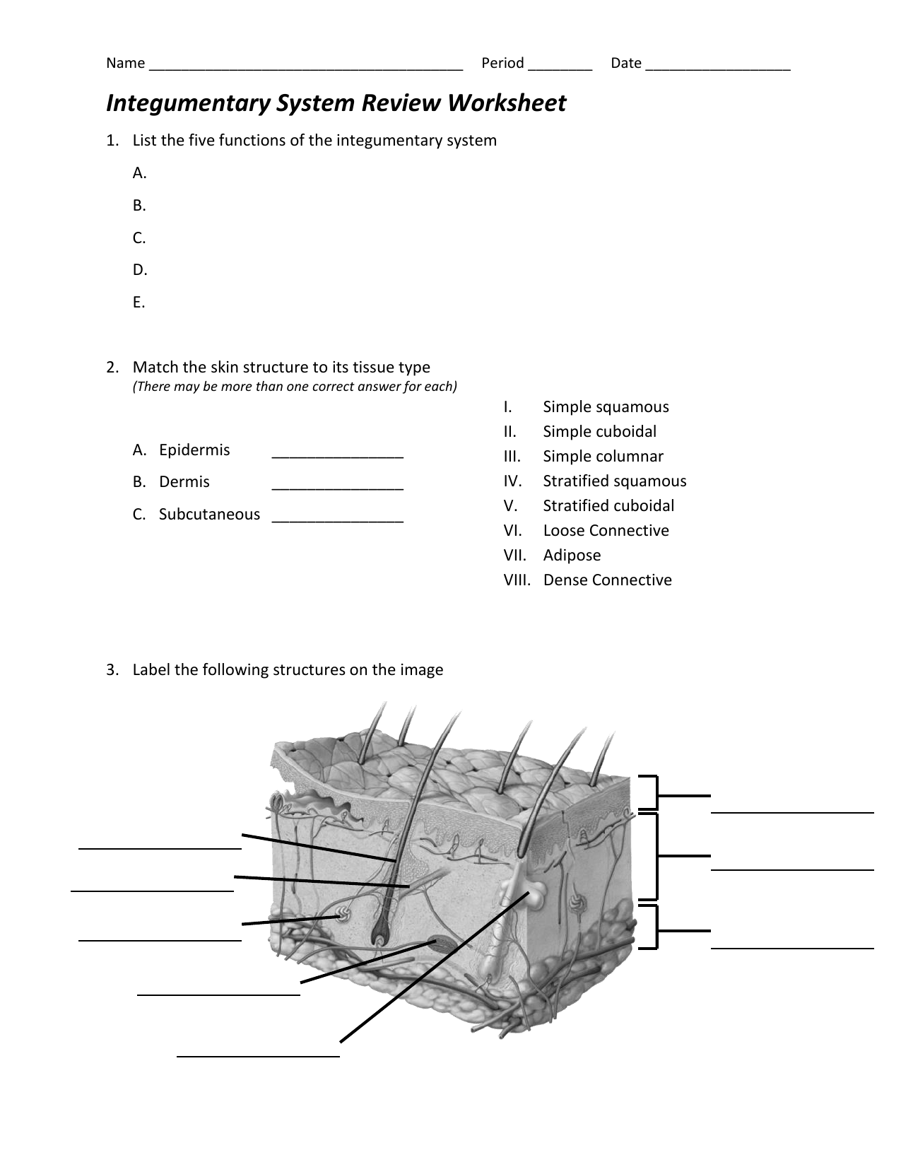
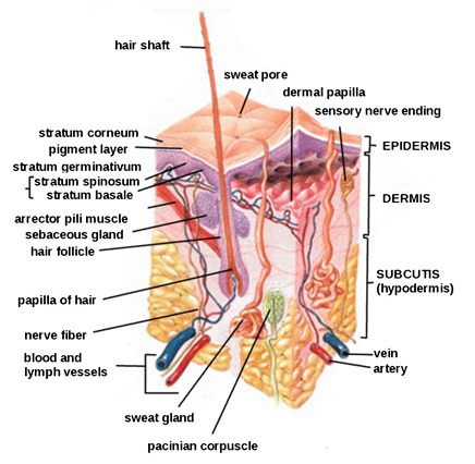
Diagram of the skin and hair includes sweat gland found in the epidermis. The integumentary system is a system full to the brim with interesting structures. Integumentary system parts the skin.
Besides the skin it comprises the hair and nails as well which are appendages of the skin. Fibers in the dermis are produced by. Contains cells that die and move to the surface.
Identify the cell type. Worksheet over the integumentary system includes diagram of the human skin to label and a series of questions about the skin. Anatomy the anatomy of the integumentary system.
Adipose cells identify the cell type. Found in the epidermis. Frizzle teaches middle school by marissa stevens.
The integumentary system or skin is the largest organ in the body. General physiology questions on human physiology. What are the names and functions of the two main layers of the skin.
0 0000 a shoutout is a way of letting people know of a game you want them to play. In humans this system accounts for about 15 percent of total body weight. However its not the only part.
Youll likely think of the skin. Then complete the statements that follow. The deeper subcutaneous stratum on the other hand is made up of connective tissue and fatty substances.
More information find this pin and more on ms. Urinary system diagram to label. Label the skin structures and areas indicated in the accompanying diagram of thin skin.
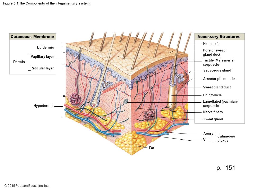
 Chapter 5 Integumentary System
2015 A P Integumentary Handout
 Skin Diagram Coloring And Labeling Worksheet Image Gallery

The Integumentary System The Integumentary System
 Integumentary System Worksheets Teaching Resources Tpt
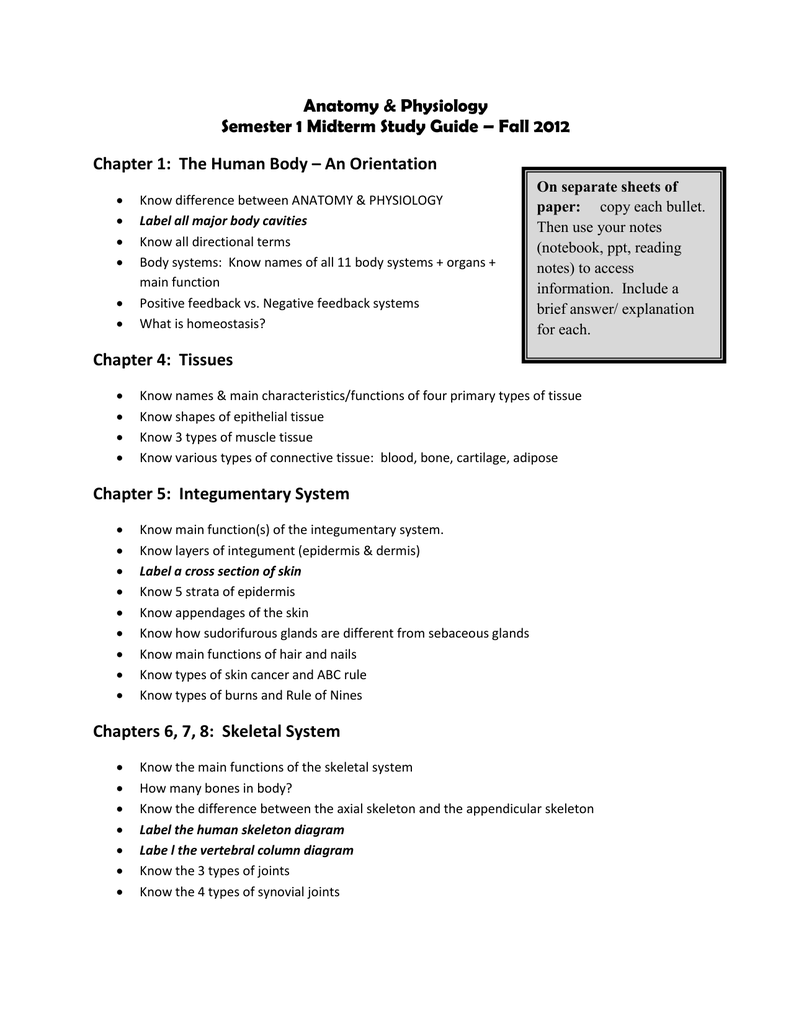
 Integumentary System Review Worksheet
 Integumentary System Biology For Majors Ii


 Anatomy Integumentary System Worksheets Teaching Resources

 Intro To The Integumentary System Packet
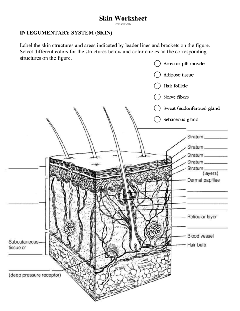 Skin Worksheet
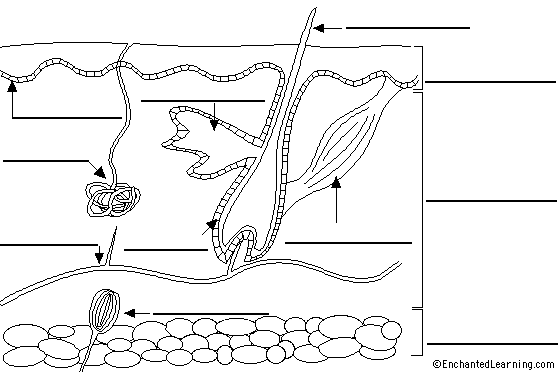 Diagram Of Skin Catalogue Of Schemas
 Integumentary System Definition Functions Organs
 Solved The Integumentary System In The Accompanying Diagr

 Integumentary System Labeling Diagram Quizlet
Imagespace Human Skin Diagram Label Gmispace Com
 Integumentary System Skin Diagram To Label Open Board
0 Response to "Integumentary System Diagram To Label"
Post a Comment